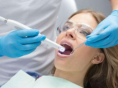
An intraoral camera is a compact, pen-sized imaging device designed to capture high-resolution, full‑color photographs inside the mouth. Unlike traditional mirror-and-probe inspections that rely only on tactile feedback and limited sightlines, the intraoral camera delivers a clear, magnified view of teeth, gums, and other soft tissues on a computer monitor. This close-up perspective makes subtle defects, early-stage wear patterns, tiny cracks, and hard-to-see decay easier to identify and document.
These cameras are built to be patient-friendly: the wand is small, ergonomically shaped, and often equipped with LED lighting to illuminate even shadowed areas. The images they produce show texture, color, and contours in ways that standard X-rays and visual exams cannot, complementing radiographs and other digital records. Because the output is immediate, clinicians can pause, zoom, and capture multiple angles in real time to ensure a complete assessment of a particular tooth or soft-tissue concern.
Beyond detection, intraoral camera images function as objective clinical evidence. High-quality photos can be added to a patient’s chart to chart progression, compare pre- and post-treatment appearances, and create a visual baseline for long-term monitoring. This objective record supports clearer communication with specialists, laboratories, and insurance reviewers when documentation is needed for clinical planning or collaborative care.
Intraoral cameras sharpen diagnostic accuracy by combining magnification with immediate visualization. When small fractures, marginal breakdown around restorations, or marginal staining are hard to see with the naked eye, the camera can reveal these issues in crisp detail. Dentists use these images alongside digital radiography and CBCT scans to form a more comprehensive clinical picture, allowing treatment plans to address both visible surface changes and underlying structural concerns.
The ability to capture and store images streamlines treatment planning. Photographs can be annotated, incorporated into digital treatment records, and compared side-by-side during case review. This is particularly valuable for restorative work, where precise assessment of existing crowns, fillings, and tooth anatomy influences material selection and margin design. Digital images also support multidisciplinary planning when coordinating care with orthodontists, periodontists, or prosthodontists.
Because images are taken noninvasively and repeated as needed, the intraoral camera is a practical tool for staging care. Clinicians can document an issue at its earliest appearance, revisit it at recall appointments, and make evidence-based decisions about when to intervene. That measured approach helps avoid premature treatment while ensuring issues do not progress unnoticed.
One of the strongest advantages of intraoral cameras is how they improve patient communication. When patients see the same magnified view as the clinician, abstract explanations become concrete. A visual demonstration of a tiny crack, a hairline defect in a filling, or early enamel breakdown helps patients understand the rationale behind recommended care, making informed decisions less stressful and more collaborative.
Images produced by the camera also empower patients to track outcomes. Whether monitoring the healing of soft tissue after a procedure, evaluating the fit of a restoration, or following whitening progress, patients who can visually confirm changes are often more engaged in homecare and more likely to adhere to follow-up recommendations. This transparency builds trust and fosters a stronger clinician–patient relationship.
For people who feel anxious about dental visits, the camera can be reassuring. It reduces the sense that findings are hidden or speculative and replaces ambiguity with clear visuals. The practice’s focus on showing, not just telling, helps patients feel respected and involved in their own care journey.
From a workflow perspective, intraoral cameras increase efficiency and precision. A quick series of images taken at the start of an exam can guide the clinician’s focus, reduce chair time spent explaining findings, and create a reliable visual log for future visits. For auxiliary staff, these images make room for consistent, standardized documentation that supports scheduling, treatment sequencing, and case acceptance discussions.
These cameras are especially helpful in collaborative cases. When referring to a specialist or sending cases to a dental laboratory, high-resolution intraoral images convey important clinical details that words alone cannot. Labs use these photographs to match shade, understand contours, and anticipate margins, while specialists rely on them to triage referrals and prepare for operative appointments.
Additionally, intraoral images support quality assurance. They provide an auditable trail of clinical conditions and procedures, which is useful for internal review and continuity of care. Because the images are reproducible, they help the team maintain consistent standards and revisit clinical decisions with clear visual evidence when needed.
An intraoral camera exam is quick and comfortable. During a routine visit, the clinician or hygienist will briefly introduce the wand and explain what they plan to photograph. The camera is moved gently along the teeth and soft tissues while live images appear on a screen; the operator may ask the patient to bite down or reposition slightly to capture optimal angles. Most image captures take only seconds each, and the entire process can be completed within the normal flow of a clean-and-check appointment.
Patients do not need any special preparation. The camera is noninvasive and uses visible light—there is no radiation exposure involved—so it is safe for nearly all patients, including those who are monitoring ongoing oral health conditions. Images are immediately reviewed with the patient; clinicians use them to explain findings, sketch treatment steps, or document observations for the patient’s record.
After images are captured, they are saved in the digital chart and can be referenced at any future visit. This makes follow-up conversations straightforward: clinicians can show previous photographs alongside current ones to illustrate changes and justify recommended actions. Because the images become part of the permanent record, they also enable precise comparisons over months and years.
Overall, the intraoral camera is a practical, patient-centered technology that supports better detection, clearer communication, and more predictable outcomes. At Studio Dental Center for Advanced Dentistry, we integrate intraoral imaging into exams to enhance clinical decision-making and to help patients understand their oral health more fully.
In summary, intraoral cameras bring a new level of detail and clarity to dental examinations—improving diagnosis, strengthening communication, and supporting coordinated care. If you have questions about how this technology is used during visits or how it might benefit your specific situation, please contact us for more information.


An intraoral camera is a small, pen-sized imaging device that captures high-resolution, full-color photographs inside the mouth. It provides magnified views of teeth, gums, and other oral tissues that are displayed on a monitor in real time. Because the wand is ergonomic and fitted with LED illumination, clinicians can document texture, color, and fine surface detail that are difficult to see with the naked eye.
These images complement traditional examinations and digital radiography by showing surface features such as cracks, marginal breakdown, and staining. The camera output can be paused, zoomed and captured from multiple angles to ensure a complete assessment of a specific area. Saved images become part of the patient record and support ongoing monitoring and clinical decision-making.
An intraoral camera captures external surface detail using visible light, while X-rays and CBCT scans reveal internal structures such as bone, root anatomy and interproximal decay. The camera excels at showing color, texture and fine surface irregularities that radiographs cannot depict, so it is an important visual complement to radiographic imaging. Each modality provides different but complementary information that together form a fuller diagnostic picture.
Unlike radiographs, intraoral images do not use ionizing radiation and can be repeated as often as needed during an exam without exposure concerns. Clinicians commonly compare photographs with radiographs and scans to confirm a diagnosis and refine a treatment plan. This multimodal approach improves accuracy and helps prioritize care based on both surface and structural findings.
Intraoral images increase diagnostic precision by magnifying subtle findings that might otherwise be missed, such as hairline fractures, marginal gaps around restorations and early enamel defects. When clinicians can see these details on a screen alongside radiographs and CBCT data, they can form a more complete understanding of the condition. Annotated photographs become a practical tool for planning restorative margins, choosing materials and sequencing care.
Because images are saved and organized in the digital chart, clinicians can compare before-and-after states and track progressive changes over time. This documentation supports staged care by helping providers decide when intervention is necessary versus when monitoring is appropriate. The ability to review, mark up and share images also streamlines case discussions with specialists and laboratory technicians.
An intraoral camera exam is typically quick and comfortable and adds only a few minutes to a standard dental visit. The wand is small and designed for gentle intraoral use, and most image captures take only seconds each to obtain optimal views. Operators will ask simple bite or head-position adjustments as needed but generally complete the process within the normal flow of a clean-and-check appointment.
Because the camera uses visible light and is noninvasive, there is no special preparation required and no recovery time after imaging. For patients with sensitive gag reflexes or limited mouth opening, clinicians can adapt technique and positioning to minimize discomfort. Overall, the additional time spent capturing images often reduces chair time later by clarifying findings immediately.
Yes. Intraoral cameras use visible LED illumination and do not emit ionizing radiation, making them safe for most patients, including children and those who require frequent monitoring. The device is noninvasive and can be used repeatedly without exposing patients to additional radiation. Clinicians can tailor imaging frequency and technique to each patient’s needs and comfort level.
For patients with special health conditions or limited cooperation, the clinical team can modify the approach or capture fewer images to maintain comfort while still documenting important findings. If a patient has specific concerns or mobility limitations, staff will explain the process and make accommodations to ensure a safe, respectful experience. The emphasis is on gentle handling and clear communication throughout the exam.
Intraoral photographs are saved directly to the patient’s digital chart where they become part of the permanent record and can be referenced at future visits. These images create a visual baseline that allows clinicians to monitor changes, verify healing and reassess restorations over time. Saved photos can be annotated and compared side-by-side to document progression or treatment outcomes.
Stored images also support clear communication with other members of the care team and with outside specialists when collaborative planning is necessary. Because photographs reproduce surface detail accurately, they help the team maintain consistent clinical standards and provide an auditable trail for quality assurance and continuity of care. At times when additional perspective is needed, these images are paired with radiographs and CBCT data for comprehensive review.
Absolutely. High-quality intraoral images help clinicians evaluate tooth anatomy, shade variation, surface texture and the fit of existing restorations—all factors that influence cosmetic and restorative planning. Photographs allow for precise discussion about margins, contour and aesthetic goals, which improves material selection and preparation design. Visual documentation also helps set realistic expectations by showing the current condition before treatment begins.
These images are commonly used to communicate detailed information to dental laboratories for shade matching and contouring of crowns, veneers and other restorations. When combined with digital impressions and radiographic data, intraoral photos contribute to predictable outcomes and smoother laboratory workflows. The result is more consistent aesthetic integration and functional performance for restorative cases.
Sharing intraoral images during an exam makes findings visible and concrete, which helps patients understand the clinical rationale behind recommendations. Seeing a magnified crack, a marginal defect or early enamel erosion reduces ambiguity and fosters informed, collaborative decision making. This transparency typically increases patient confidence in recommended care and encourages adherence to follow-up and homecare instructions.
Visual evidence also supports discussions about treatment urgency and monitoring strategies, allowing patients to weigh options with clearer context. When patients can review, ask questions and view annotated images, consent conversations are more thorough and focused. The approach helps build a stronger clinician–patient relationship based on mutual understanding and respect.
Dental teams routinely include intraoral photographs when referring a case to a specialist or sending instructions to a dental laboratory because images convey surface detail that written descriptions alone cannot. Labs use photos to match shade and anticipate margin contours, while specialists rely on them to triage referrals and plan operative strategies. High-resolution images reduce the need for excessive follow-up questions and support more efficient treatment coordination.
When images accompany radiographs and digital scans, they create a richer dataset for collaborative planning and help ensure all clinicians involved see the same clinical picture. This alignment reduces surprises at the time of treatment and contributes to more predictable, higher-quality outcomes. Stored photographs also make it easier to revisit decisions and maintain continuity when multiple providers are involved in long-term care.
During an intraoral camera exam the clinician or hygienist will briefly explain the process, gently move the wand around the teeth and soft tissues, and display live images on a monitor for review. Captures typically take only seconds each and require simple repositioning or biting adjustments to obtain ideal angles. The procedure is noninvasive, uses visible light, and adds minimal time to a routine visit.
After images are captured they will be saved to your digital chart and reviewed with you so you can see the same views as the clinician. These photographs are then used to document current conditions, support treatment planning and track changes at future visits. If you have questions about what is shown, the team will take time to explain findings and next steps in plain language.
Ready to book your next dental visit or have questions about your care?
At Studio Dental Center for Advanced Dentistry, our team is here to make getting started simple and stress-free. Whether you call, email, or submit our online form, we’re happy to help with scheduling, treatment questions, or anything you need along the way. We take the time to listen and guide you through your options so you feel confident and informed at every step. Take the first step toward a healthier, more confident smile, reach out today and experience personalized care made easy.