At Studio Dental Center for Advanced Dentistry, we combine experienced clinical judgment with advanced imaging to give patients clearer answers and more predictable results. Cone-beam computed tomography (CBCT) is one of the technologies we rely on to visualize anatomy that cannot be fully captured with traditional two-dimensional X-rays. By producing high-resolution, three-dimensional images of the teeth, jaws, airway, and surrounding structures, CBCT helps our team understand the whole picture before recommending treatment.
CBCT is not a separate specialty—it’s a diagnostic tool that supports many aspects of modern dentistry. Used thoughtfully, it accelerates diagnosis, improves treatment planning, and reduces uncertainty for both clinicians and patients. The result is care that is better informed, more efficient, and tailored to each patient’s unique anatomy and goals.
Traditional dental X-rays are excellent for many routine needs, but they compress complex anatomy into flat images. CBCT captures volumetric data, allowing clinicians to examine structures from multiple angles and at varying depths. This three-dimensional perspective exposes relationships and details—such as root morphology, bone volume, and tooth position—that can be obscured in two-dimensional films.
With CBCT, clinicians can identify issues that might otherwise be missed or misinterpreted, including hidden canals, tiny fractures, or proximity to critical anatomic landmarks like the inferior alveolar nerve or maxillary sinus. This enhanced visibility supports more accurate diagnoses and helps avoid surprises during treatment.
Importantly, CBCT scans are localized and targeted for dental use. The images are captured quickly, and viewing software allows the team to manipulate slices and reconstructions to focus on the areas that matter most for a given case. That flexibility makes CBCT a practical and powerful addition to the diagnostic toolkit.
Effective treatment begins with a clear plan. CBCT makes planning more precise by providing accurate measurements of bone height, width, and density, as well as spatial relationships between teeth and surrounding anatomy. Whether preparing for restorative work, endodontics, or surgical procedures, these measurements help the clinician select appropriate techniques and anticipate technical challenges.
For complex restorative cases, CBCT data can be integrated with digital scans and planning software to visualize outcomes and simulate steps in a treatment plan. This coordinated workflow reduces guesswork and supports predictable results—especially when multiple disciplines (restorative, periodontal, surgical) need to work together.
The ability to review a case three-dimensionally also improves patient communication. When patients can see their anatomy rendered in 3D and understand the rationale behind recommended treatments, they are better positioned to make informed decisions about their care.
Dental implant therapy benefits directly from the spatial accuracy of CBCT imaging. Successful implant placement depends on precise knowledge of available bone, the location of nerves and sinuses, and the ideal orientation for each implant. CBCT allows clinicians to plan implant positions virtually, minimizing risk and maximizing functional and aesthetic outcomes.
In surgical cases, CBCT helps with preoperative mapping and intraoperative guidance. Surgeons can anticipate variations in anatomy, select appropriate implant sizes, and, when indicated, design surgical guides that translate the virtual plan to the clinical setting. This advance preparation reduces intraoperative uncertainty and enhances procedural efficiency.
Beyond implants, CBCT is useful for evaluating impacted teeth, assessing pathology, and guiding complex extractions. For cases that involve the jaw joints or airway, CBCT offers objective data that informs both diagnosis and long-term management strategies.
Patient safety is a primary consideration with any imaging technology. Dental CBCT systems are designed specifically for head and neck imaging and typically use lower radiation doses than conventional medical CT scans. Modern units and appropriate imaging protocols allow clinicians to limit the field of view and exposure to what is essential for the clinical question at hand.
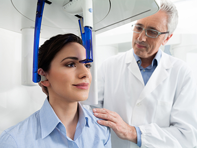
The scan itself is quick—often completed in under a minute—and the patient sits or stands comfortably while the machine rotates. Because CBCT captures the entire region of interest in a single pass, repeat exposures are usually unnecessary, and the overall patient experience is efficient and noninvasive.
Our team follows evidence-based guidelines to determine when CBCT is clinically justified. We weigh the diagnostic benefit against exposure and use the smallest effective field of view to answer the specific question. This measured approach helps ensure patient safety while providing the best possible information for treatment planning.
CBCT is a versatile tool that supports a wide range of dental services, from routine assessments to specialized treatments. In everyday practice it can clarify ambiguous findings on conventional films, support endodontic decision-making, and inform periodontal assessments. In specialty areas such as oral surgery, implantology, and orthodontics, CBCT often plays a central role in planning and execution.
Because CBCT files can be exported and shared in standard formats, they facilitate collaboration with other specialists and labs. This interoperability streamlines coordinated care—especially for multidisciplinary cases where precise alignment between teams is critical for success.
Alongside clinical benefits, CBCT contributes to a safer, more predictable care pathway. By reducing uncertainty and enabling targeted interventions, it helps minimize unnecessary procedures while improving outcomes. When used judiciously, CBCT enhances the quality of care across a broad spectrum of dental needs.
In summary, cone-beam computed tomography is a powerful diagnostic tool that brings three-dimensional clarity to dental care. At Studio Dental Center for Advanced Dentistry, we use CBCT to improve diagnostic confidence, support precise treatment planning, and enhance patient safety and comfort. If you’d like to learn more about how CBCT may play a role in your dental care, please contact us for more information.


Cone-beam computed tomography, commonly called CBCT, is a three-dimensional imaging technology designed for dental and maxillofacial use. The system captures a volumetric dataset by rotating a cone-shaped X-ray beam around the head, producing high-resolution images of teeth, bone, and surrounding anatomy. Unlike traditional two-dimensional films, CBCT renders cross-sectional views and volumetric reconstructions that reveal spatial relationships and internal structures.
CBCT is a diagnostic tool rather than a treatment in itself and is used to answer specific clinical questions that cannot be resolved with standard X-rays. The scanner can be set to focus on a limited field of view so clinicians obtain only the region needed for diagnosis. Clinicians review the images using dedicated software to evaluate anatomy from multiple angles and plan care accordingly.
Conventional dental X-rays produce flat, two-dimensional images that compress complex anatomy onto a single plane, which can obscure depth and spatial relationships. CBCT acquires volumetric data that can be viewed as axial, sagittal, coronal slices or as three-dimensional reconstructions, revealing details such as root morphology, bone contours, and the relative positions of anatomic landmarks. This three-dimensional perspective reduces ambiguity and helps clinicians distinguish overlapping structures that may be indistinct on 2D films.
CBCT systems used in dentistry are optimized for high spatial resolution of hard tissues and for limited fields of view, making them more targeted than medical CT scans. While CBCT often involves a higher dose than a single periapical film, modern units and protocols aim to minimize exposure by restricting the scanned area to what is clinically necessary. The choice between 2D and 3D imaging depends on the diagnostic question and the clinician's judgment.
CBCT is valuable in implant planning, complex endodontics, oral and maxillofacial surgery, orthodontics, and evaluation of the airway and temporomandibular joints. For implants, it helps assess bone volume and proximity to nerves and sinuses; for endodontics, it can reveal hidden canals, root fractures, or periapical pathology that are not visible on 2D films. Surgeons also use CBCT to evaluate impacted teeth, lesions, and bony anatomy prior to procedures.
Orthodontists and airway specialists may use CBCT when three-dimensional relationships influence treatment decisions, such as assessing airway volume or craniofacial asymmetry. Because CBCT files can be exported in standard formats, they support collaboration with labs and specialists for multidisciplinary cases. Ultimately, CBCT is used when the added diagnostic value justifies the targeted exposure.
CBCT provides precise measurements of bone height, width, and density, and it shows the spatial relationship between proposed implant sites and critical structures like the inferior alveolar nerve and maxillary sinus. These data allow clinicians to virtually position implants, choose appropriate diameters and lengths, and assess the need for bone grafting or sinus augmentation before entering the operatory. Virtual planning can also be combined with intraoral scans to design prosthetically driven implant positions that optimize function and esthetics.
When indicated, CBCT-based plans can be translated into surgical guides that help reproduce the virtual implant trajectory intraoperatively, reducing guesswork and improving accuracy. This coordinated digital workflow supports predictable outcomes and helps teams anticipate restorative and surgical challenges ahead of time. The result is a more controlled treatment pathway with clearer expectations for both clinician and patient.
CBCT systems used in dentistry are engineered specifically for head and neck imaging and generally use lower radiation doses than conventional medical CT scanners, though doses vary by unit, field of view, and exposure settings. Clinicians apply the principle of ALARA, or as low as reasonably achievable, selecting the smallest effective field of view and the lowest acceptable exposure for the clinical task. When a CBCT scan is clinically justified, the diagnostic benefit is weighed against the exposure to ensure patient safety.
Modern CBCT protocols and equipment features, such as adjustable voxel size and collimation, help limit dose while preserving image quality for the question at hand. Lead shielding and standard safety procedures further reduce unnecessary exposure during imaging. If you have specific concerns about radiation, discuss them with the clinical team so they can explain the rationale and safety measures used for your case.
A CBCT scan is typically quick and noninvasive, often completed in under a minute once the patient is positioned. You will be asked to sit or stand still while the machine rotates around your head and captures the images; remaining motionless is important to avoid motion artifacts. Metal objects such as jewelry, eyeglasses, removable dental appliances, or hairpins should be removed from the region being imaged to prevent image distortion.
The process is generally comfortable, with no need for injections or contrast agents for routine dental CBCT scans. After acquisition, the images are reconstructed and reviewed by the clinician using specialized software to answer the clinical question and plan treatment. The team will explain the findings and how the images inform your care during your consultation.
CBCT has limitations including reduced soft-tissue contrast compared with medical CT and susceptibility to artifacts from metal restorations, orthodontic appliances, or patient motion. These artifacts can obscure detail in the region of interest and sometimes necessitate alternative imaging approaches. In cases where soft-tissue characterization is essential or when very large fields of view are required, a medical CT or MRI may be more appropriate.
Pediatric patients and pregnant patients warrant careful consideration, and clinicians will avoid imaging when the diagnostic yield does not justify exposure. Additionally, very small or subtle findings may still require complementary imaging or clinical correlation to provide a complete assessment. The decision to use CBCT is always based on clinical indication and the expected value of the information obtained.
Clinicians use CBCT data to obtain accurate anatomic measurements, identify variations that affect treatment, and simulate procedures before performing them on patients. In endodontics, CBCT can reveal complex canal anatomy or root fractures that change the treatment approach; in surgery, it supports preoperative mapping and the creation of surgical guides. Integrating CBCT with digital impressions and planning software enables a coordinated workflow that aligns surgical and restorative goals.
CBCT also enhances communication among multidisciplinary teams and with patients by providing clear visualizations of anatomy and planned interventions. Sharing standardized DICOM files facilitates collaboration with specialists and laboratories, reducing misunderstandings and supporting consistent execution of the plan. The overall effect is more informed decision-making and better alignment between expected and achieved outcomes.
Preparation for a dental CBCT scan is minimal and typically involves removing jewelry, eyeglasses, hairpins, and removable dental appliances that could interfere with the image. Wear comfortable clothing and avoid metal accessories around the head and neck area when possible, as these can produce artifacts. If you are pregnant or suspect you might be, inform the dental team so they can assess whether imaging is appropriate and consider alternative approaches if necessary.
If you have prior imaging or treatment records relevant to the region being evaluated, bring them or notify the office so clinicians can compare studies. The staff will explain positioning and what to expect during the scan and will answer any safety questions before acquisition. Clear communication about medical history and current symptoms helps the team select the most appropriate imaging protocol.
At Studio Dental Center for Advanced Dentistry, CBCT is used as part of a comprehensive diagnostic and planning process that supports individualized treatment recommendations. The technology is combined with clinical examination and, when appropriate, digital impressions or other imaging to create a coordinated plan that addresses restorative, surgical, and periodontal considerations. Images are reviewed with patients to explain findings and rationale for recommended next steps in a clear, visual way.
The team follows evidence-based guidelines to determine when a CBCT scan is justified and selects protocols that limit exposure while providing the necessary information. When multidisciplinary input is needed, CBCT datasets are shared with specialists and labs in standard formats to streamline collaboration. This measured use of advanced imaging helps enhance diagnostic confidence and support predictable, patient-centered care.

Ready to book your next dental visit or have questions about your care?
At Studio Dental Center for Advanced Dentistry, our team is here to make getting started simple and stress-free. Whether you call, email, or submit our online form, we’re happy to help with scheduling, treatment questions, or anything you need along the way. We take the time to listen and guide you through your options so you feel confident and informed at every step. Take the first step toward a healthier, more confident smile, reach out today and experience personalized care made easy.